This is Clarus Viewer®
Revolutionizing medical imagery through artificial intelligence
Key Features
Imports MRI and CT DICOM anatomical datasets
Allows models to be rotated, scaled, measured, annotated, sliced, and cut to observe and evaluate interior structure, tissue and fluids
Provides ability to adjust visual features of images/models at user’s discretion
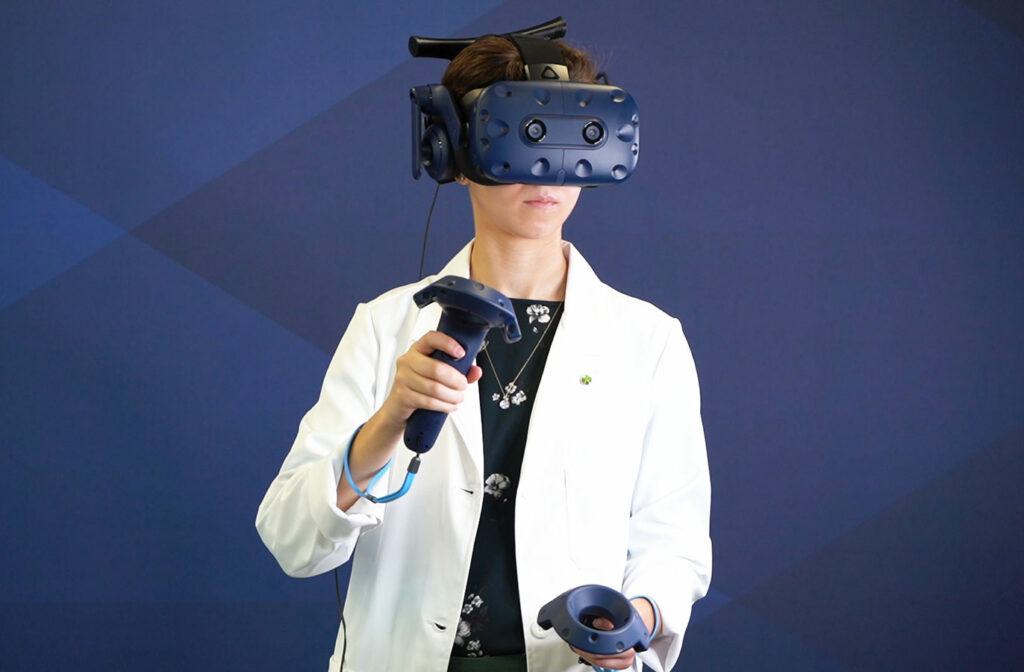
Digital Solutions
Scan imagery for some patients with complex cases or irregular anatomy is difficult to analyze and evaluate with traditional 2D viewers.
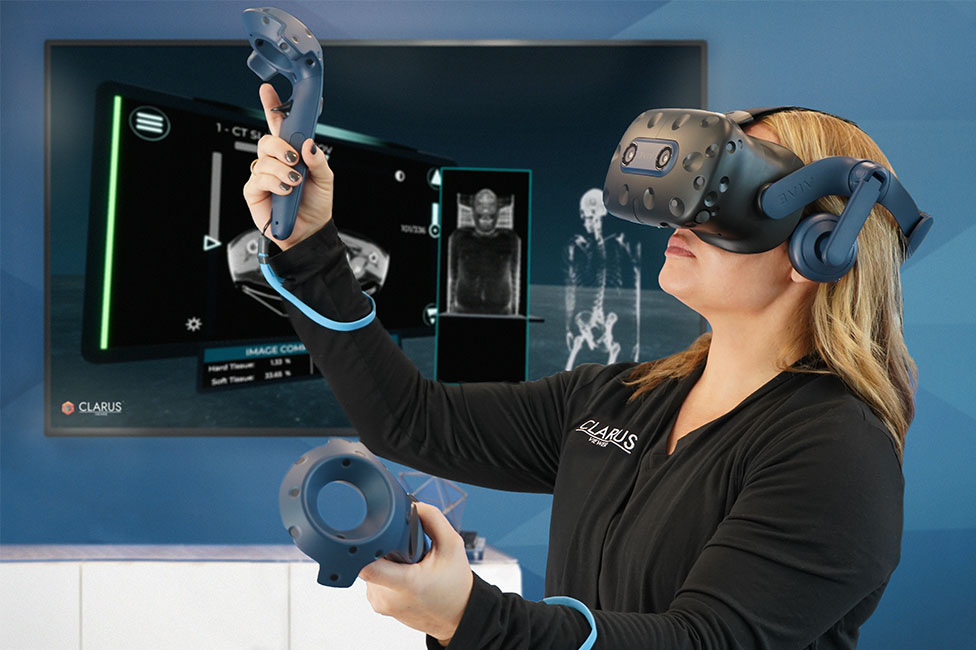
Clarus Viewer is a software solution created to allow users to visualize and manipulate 2D images and 3D models of patients in Virtual Reality (VR).
The innovative methods of Clarus Viewer for medical image visualization and collaborative analysis advance Digital Healthcare using technologies from Digital Engineering.
Clarus Nexus™ coming soon!
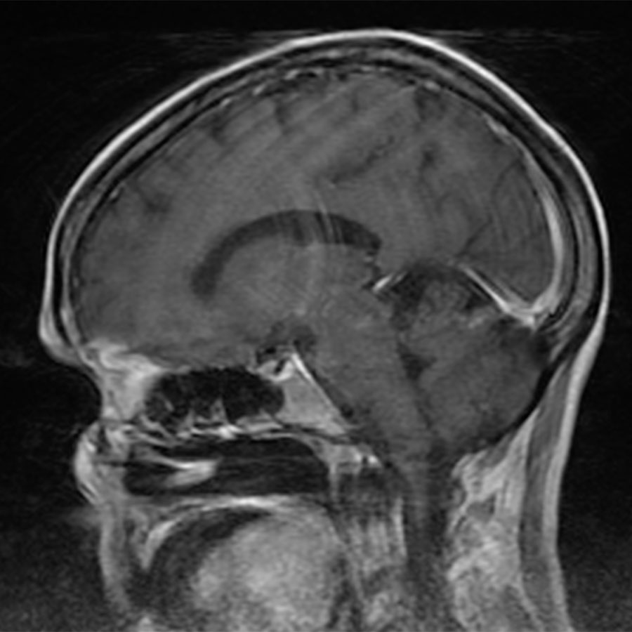
Sample DICOM images for import
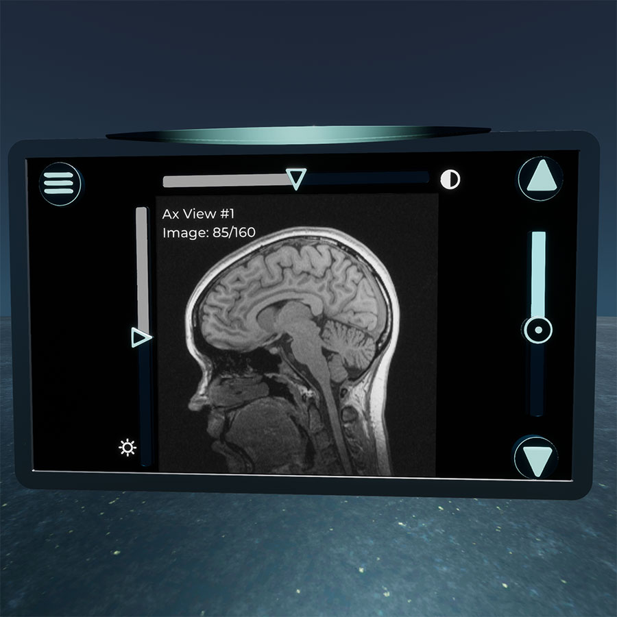
Virtual 2D image Scan Deck
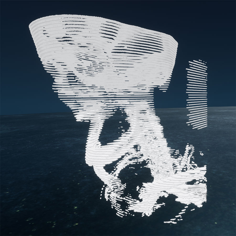
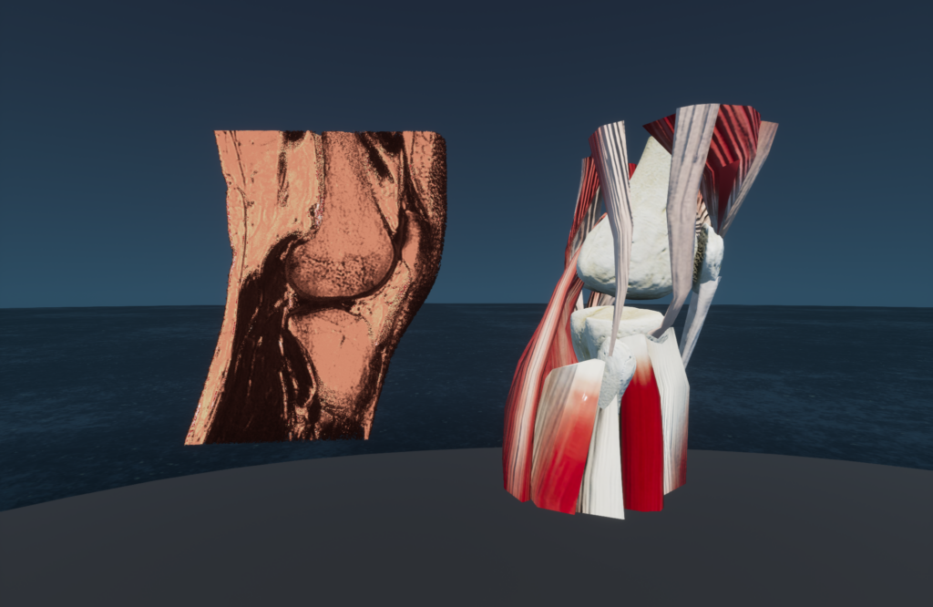
Virtual 3D Voxel Mesh: Hard Tissues
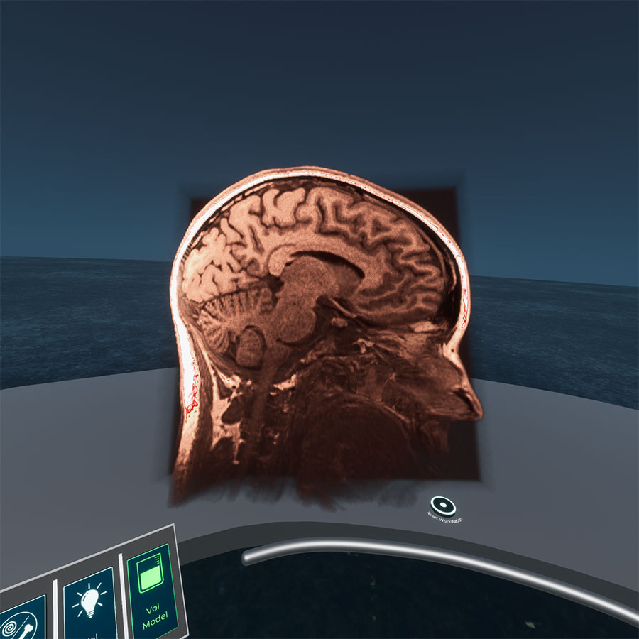
Virtual 3.5D Volumetric Mesh: Soft/Hard Tissues
We're Different
Clarus Viewer is different from the status quo and existing viewers because it presents 2D and innovative 3D models that can be intuitively manipulated and analyzed in ways not possible with today’s scan readers using the below features:
- 2D and 3D Visualization in Virtual Reality
- 3D Scaling, Slicing, Measuring and Annotation
- Anonymized Patient Data

Collaboration
Clarus Viewer Corporation and the University of Alabama in Huntsville (UAH) College of Nursing are excited to announce its collaboration in advancing medical technologies.
Advancing Education
Clarus Viewer Corporation is thrilled to announce the completion a new virtual reality (VR) educational application – Clarus Viewer TrainerTM . Clarus Viewer Trainer is a training aid for healthcare students and instructors focused on bridging the gap between traditional book learning and patient care.
Contact Us
Want to know more about Clarus Viewer?
Please feel free to reach out to the Clarus Viewer Lead Market Developer and Medical Liaison, Cayla Garrett, BSN, RN.

Clarus Viewer Corporation™
5030 Bradford Drive, NW; Building 2, Suite 104,
Huntsville, AL 35805
Clarus Viewer® v1.0 is a 510(k) cleared medical device.
